

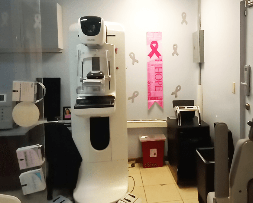
At Imaging Center PC in St. Croix Virgin Islands, our Hologic equipment does 3D, 2D, and specimen mammography.
3D mammograms are a stack of computer generated 1-millimeter-thick images (aka slices/tomograms) through every breast level. Camera movement information produces the 3D slices. Mammo-Radiologists manipulate these files in three dimensions to subtle abnormalities in dense breasts. We use 2D, synthetic 2D, and/or 3D in our digital mammography protocols.
Our team commits to providing personalized care for screening, diagnostic, and interventional services. We prioritize your comfort and safety.
Occasionally, screened patients are called back because:
- It is necessary for a sonographer to examine their very dense breast(s).
- Additional mammographic imaging can prove a screen finding is: a. An artifact. b.Benign. c. Probably benign & needs a 6-month follow-up.
- The concern for malignancy suffices to recommend a biopsy. Most biopsy results are benign, but some reveal cancer or precancerous conditions.
- Schedule your mammogram during a time of the month when your breasts are least tender, usually shortly after your period.
- Bring prior mammogram report and DICOM digital images to us the day of your new mammogram or, before the appointment, ask your prior mammography facility to send the report and digital images to us.
- Before your mammogram, mention every breast implant that you have to an ICPC receptionist and mammographer.
- Wear pants or a skirt so you can stay partially dressed throughout the procedure.
- Consider taking pain relievers ½ hour before the mammogram if compression of breast is very uncomfortable.
- Relax to reduce tension and improve image quality.
- Hold your breath when told to do so.
- Use deodorant, powder, or lotion on your breasts.
- Wear bulky jewelry. Leave it at home.
A mammogram is an X-ray image of the breast used to detect changes in breast tissue, such as lumps, cysts, or calcifications, that could indicate cancer. It is a vital tool in early breast cancer detection, allowing physicians to identify the disease at its most treatable stages. Mammograms are categorized into screening mammograms, used for routine checkups in women without symptoms, and diagnostic mammograms, which are used when there is a specific concern or abnormal finding that needs further evaluation.
The raw 3D mammography data is the 1mm thick 2D slices of the breast. When we keep 1mm thick slices in their z-space (3rd dimension), we see the shape of objects at different levels. Compressing this 3D volume of data into a single image results in a synthetic 2D image.
A 2d image gives detailed x-axis information, y-axis information and summated z-axis information. Because of this summation, a 2D image may show a “mass” that is not present. The 3D expansion of that 2D picture may show it to be a normal above another normal structure. The mammography camera swings when it makes a 3D mammogram. This depth discrimination allows for a clearer depiction of masses and for smaller abnormalities to be seen in dense breasts. This reduces the likelihood of callbacks for further testing.
Yes, a 3D mammogram is generally more effective in detecting early-stage breast cancer, particularly for women with dense breast tissue. The detailed 3D images allow radiologists to view the breast tissue layer by layer, which increases the chances of spotting smaller tumors or abnormalities that might not be visible in a standard 2D mammogram. This can lead to earlier diagnosis, improving treatment outcomes and overall prognosis.
Breast compression for proper mammography may cause some discomfort. When this does occur it is usually only during the brief compression. Brief breast compression is necessary to produce a sharper image with less radiation. To minimize discomfort, schedule your mammogram during the time of the month when breasts are least tender, usually a few days after your menstruation starts. Please let the technician know if you have breast tenderness so she can adjust the compression for your comfort.
The benefits of early cancer detection with mammography far outweighs the minimal risks from doing the procedure. Digital mammography radiation exposure is very low and is the same amount of radiation exposure a person gets from the natural environment in a a few months. Mammograms result in the need for additional testing and false-positive results.
During the procedure, the mammographer positions your breast on a flat platform. The mammographer gently flattens your breast using a compression paddle. This allows the radiologist to obtain a clear and accurate image. The full procedure typically takes about 15 minutes. During a 3D mammogram, the machine swings while capturing multiple images. For the best mammogram, hold your breath and do not move during the exposures. The 2D mammography is a less than 1 second breath hold per view. 3D mammography is typically four breath holds, with each lasting 4 to 5 seconds. The 3D x-ray dose is about the same as the 2D dose. After your mammogram, you can return to your normal activities.
Detecting an abnormality on your mammogram doesn't mean you have breast cancer. It could be a benign condition such as a cyst or dense tissue. You may need to undergo additional tests, such as an ultrasound or a biopsy, to further investigate the area of concern. Your radiologist will explain the results and guide you through the next steps, ensuring you understand what additional screenings or tests may be necessary.
Healthy women 40-years and older who have an average risk for getting breast cancer should have a screening mammogram every 1 to 2 years. If you have a higher risk of breast cancer because of genetic factors, personal history, and/or family history, your doctor may suggest more frequent screenings or starting earlier. Consult with your healthcare provider to determine the best breast cancer screening schedule for you.
